
Fig. 2.
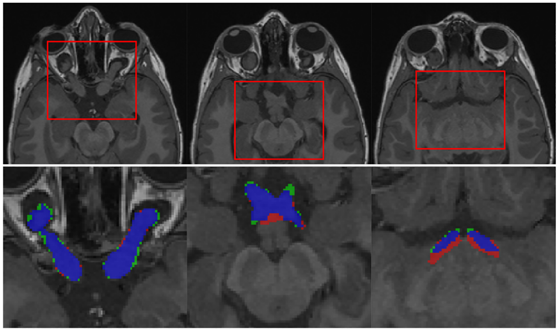
Qualitative results of AVP segmentation: selected case has large OPGs (AVP volume=5.095 ml, DSC=0.877). From left to right: 3 parts of AVP (optic nerves, chiasm and optic tracts) shown on different slices of T1 sequence. Labels: segmentation-red, ground truth-green, overlap-blue.