. 2024 Jul 24;12(7):e009550. doi: 10.1136/jitc-2024-009550

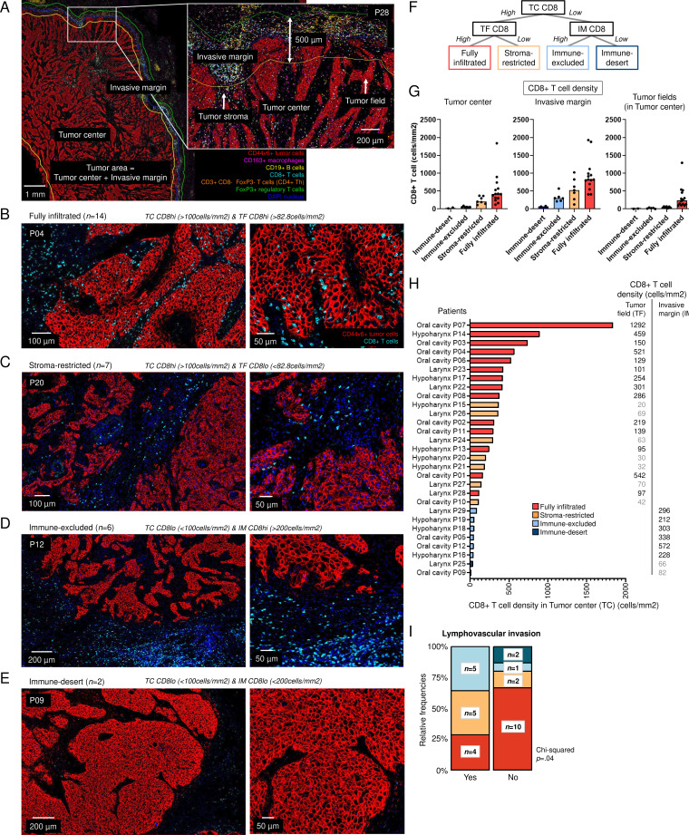
Figure 2. Immunotypes across 29 human papillomavirus-negative head and neck squamous cell carcinoma (HNSCC) resection specimens. (A) Representative image of tumor P28 with tumor center, invasive margin, tumor fields and tumor stroma. The following cells could be distinguished: CD44v6+tumor cells, CD163+macrophages, CD19+B cells, CD8+T cells, CD3+CD8 T cells (CD4+T helper cells) and FoxP3+regulatory T cells. (B–E) Representative images of tumors with (B) fully infiltrated (P04), (C) stroma-restricted (P20), (D) immune-excluded (P12), and (E) immune-desert (P09) immunotype. (F) Decision tree for assigning immunotypes and immunotypes based on CD8+T cell density in tumor center, invasive margin and tumor fields. (G) CD8+T cell densities (cells/mm2, y-axis) across immune-desert, immune-excluded, stroma-restricted and fully infiltrated HNSCC (x-axis). (H) 29 HNSCC resection specimens were assigned as infiltrated when the density of CD8+T cells in the tumor center (TC, x-axis) was higher than 100 cells/mm2. If the density of CD8+T cells in the tumor field (TF) was higher than the median density of 82.8 cells/mm2, tumor were assigned as fully infiltrated (red) and when lower than 82.8 cells/mm2, as stroma-restricted (orange). If the density was lower than 100 cells/mm2 in the tumor center but higher than 200 cells/mm2 in the invasive margin (IM), tumors were defined as immune-excluded (light blue) and when lower than 200 cells/mm2, as immune-desert (dark blue). (I) Relative frequencies and number of specimens per immunotype with the presence of lymphovascular invasion. Χ2 test was performed to obtain p value. Fully infiltrated immunotype compared with other immunotypes since groups were too small.

Figure 2