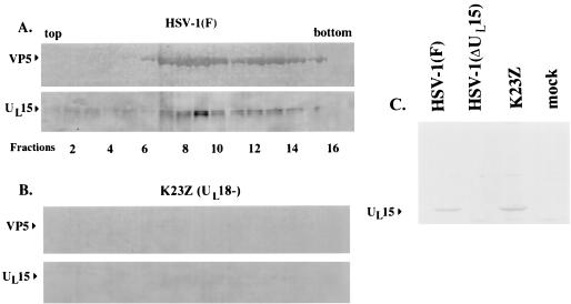
FIG. 2.
Scanned digital images of immunoblots probed with anti-UL15-MBP antibody and the NC1 antibody. Fractions (0.5 ml) of 14-ml continuous sucrose gradients containing nuclear lysates from Vero cells infected with HSV-1(F) (A) and K23Z (UL18−) (B) were collected starting at the top of the tubes. Material was electrophoretically separated, transferred to nitrocellulose, and reacted with an antibody specific for the UL15-encoded protein, UL15-MBP antiserum, and with an antibody specific for VP5, NC1. Bound immunoglobulin was visualized by addition of alkaline phosphatase-conjugated anti-rabbit antibody followed by fixation of the colored substrate. Fractions 1 to 16 are shown. (C) As a control, proteins in lysates of cells that were mock infected or infected with the indicated viruses were electrophoretically separated and reacted with UL15-MBP antiserum as indicated in panels A and B.

