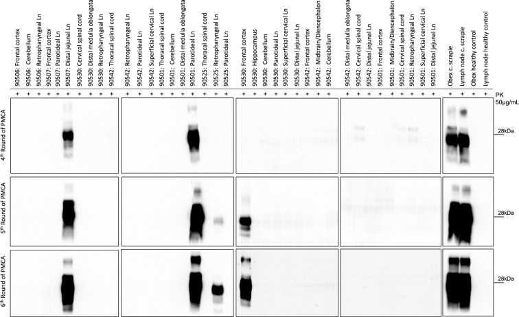
Figure 3.
Analysis of brain, spinal cord and lymph node homogenates by protein misfolding cyclic amplification (PMCA). Western blots of the 4th-6th PMCA rounds are shown. After 6 rounds of amplification, PrPSc was detected with the monoclonal antibody 6D11 in 5 out of 65 samples acquired from the inoculated sheep. Thirty-eight of these samples are shown here. Samples from healthy controls and from sheep subjected to classical scrapie were used as controls. All amplified PrPSc were characterized by a predominant di-glycosylated band. The numbers on the right of each WB indicate the molecular weight marker in kilodaltons (kDa). The amplified sample from the thoracic spinal cord of sheep 90506 is not shown but can be seen in Additional file 6, along with the remaining wells that are not included in this figure. Ln: lymph node. PK: proteinase K. C. Scrapie: classical scrapie.

