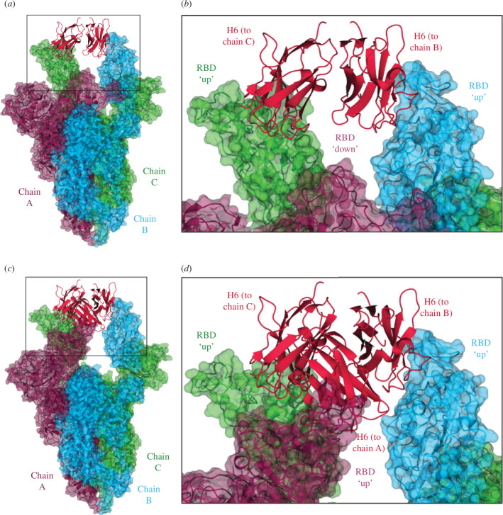
Figure 2.
EM data of spike–Nb complexes. (a) EM structure of stabilized hexaPro Omicron BA.1 spike trimer in the two RBD ‘up’ and one RBD ‘down’ conformation with two H6 nanobodies bound. H6 monomers are shown in crimson, and the spike monomers are coloured dark purple (chain A), sky blue (chain B) and lime green (chain C), with chain A in the RBD ‘down’ conformation, and chains B and C in the RBD ‘up’ conformation. (b) Close-up of the boxed region from (a) showing the binding of the H6 nanobodies to the ‘up’ RBDs of chains B and C, with no nanobody bound to the ‘down’ RBD of chain A. (c) EM structure of stabilized hexaPro Omicron BA.1 spike trimer in the three RBD ‘up’ conformation with three H6 nanobodies bound. (d) Close-up of the boxed region from (a) showing the binding of an H6 monomer to the ‘up’ RBD of each chain. Figures were generated using CCP4mg [28].

