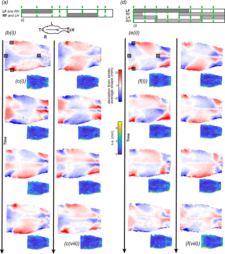
Figure 3.
Changes in equine back shape in trot (a–c) and walk (d–f). (a,d) We measured back shape (elevation) across eight instances throughout the stride (green dashed lines). Instances of the stride cycle were defined by the footfall patterns of the left and right forelimbs in trot, or the left forelimb in walk. The presentation of hindlimb timings is approximate. (b,e) Dorsal views of the equine back colour-coded by change in elevation relative to the stride-average shape at eight instances in trot (b) and walk (e); instances (i–iv) are the first column, and instances (v–viii) are the second column. Comparing across columns within a gait, pairs of stride instances are offset by half of the stride cycle and form approximate mirror images of one another, reflected about the spine. (c,f) Changes in back elevation were consistent across multiple strides, as demonstrated by the s.e.m., colour-coded across the surface. Typical errors were an order of magnitude less than the peak changes in elevation. The error presented captures errors in identifying the same instances in time, aligning or registering the shapes and measurement errors. Each instant represents the average and s.e. in elevation over (b,c) 6–8 strides (n = (6,7,7,6,7,8,7,8)) at trot, and (e,f) 5–6 strides (n = (5,5,5,6,5,5,5,5)) at walk. (a) LF: left forelimb, RH: right hindlimb; RF: right forelimb and LH: left hindlimb. (a) Inset of dorsal view of horse displaying H: head, T: tail, L: left, R: right. Grey squares represent motion-capture markers placed at (in cranio-caudal direction): T6, tuber coxae (two markers) and Cd1.

