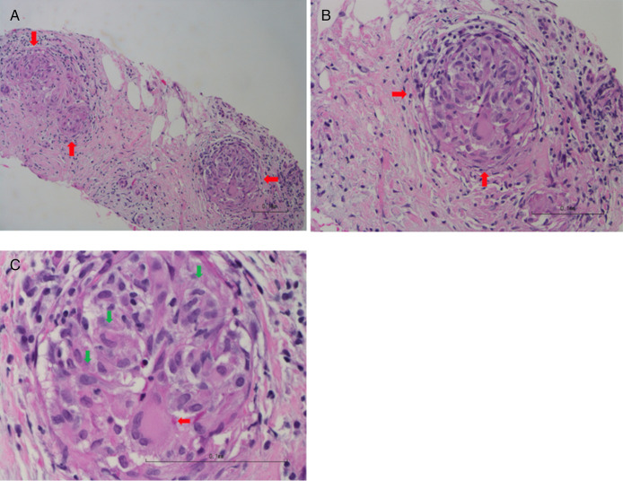
Figure 2.
(A) Low power (10×) of the pancreatic fine needle aspiration cell block has denuded pancreatic acini and ducts. Three granulomas are seen (red arrows). (B) Medium power (20×) highlights lymphocytes on the right side with a granuloma (red arrows). (C) Higher power (40×) highlights 1 granuloma composed of epithelioid macrophages (green arrows) and multinucleated giant cells (red arrows).

