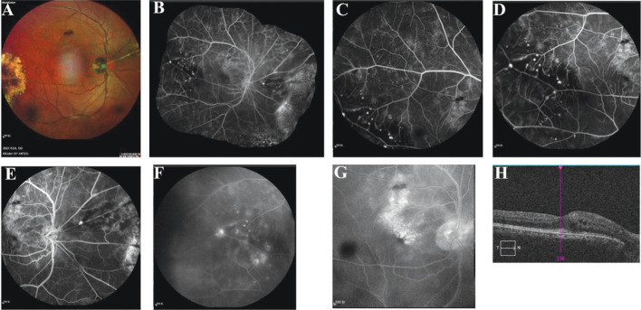
Figure 2. Multimodal imaging data of a patient with Coats disease (No.9).
A: Right nasal artery of the right eye featured silvery changes, aneurysmal enlargement was present on the superior nasal artery surrounded by exudation and small pieces of hemorrhage, flaky hemorrhage was present in the superior temporal part of the retina, and large yellowish-white exudation was observed in the temporal periphery; B: Puzzle of the fundus image of the right eye; C: Right temporal peripheral capillary dilatation; D: Focal tumor-like hyperfluorescence with a capillary nonperfusion area in the peripheral arteriolar end of the right temporal eye; E: Focal tumor-like hyperfluorescence on the main trunk of the lateral nasal artery of the right eye; F: Fluorescence leakage in the corresponding focal area with a prolonged contrast time; G: Cystic fluorescence accumulation in the arch ring area in the late stage; H: Cystic edema in the macular area was observed on OCT. OCT: Optical coherence tomography.

