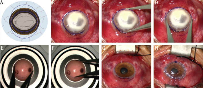
Figure 1. The diagram and surgical procedures of FPK.
A, B: Picture A is the diagram of picture B. A suitable trephine (the yellow circle) is larger than the longest diameter of the ulcer. Two methylene blue marks (the solid lines of the two blue circle) were made with the ulcer included inside using the trephine, and the intersection of the two arcs was shown in a fusiform shape. C, D: The Castroviejo circle gauge was used to measure the longest and the shortest diameter of the fusiform recipient bed. E, F: The Castroviejo circle gauge was used to mark the longest and the shortest diameter of the graft, then a fusiform graft was cut along the indentation using corneal scissors. G: The diseased cornea was dissected along the indentation of the recipient bed. H: The donor cornea was sutured to the recipient bed with 16 interrupted 10/0 nylon sutures. FPK: Fusiform penetrating keratoplasty.

