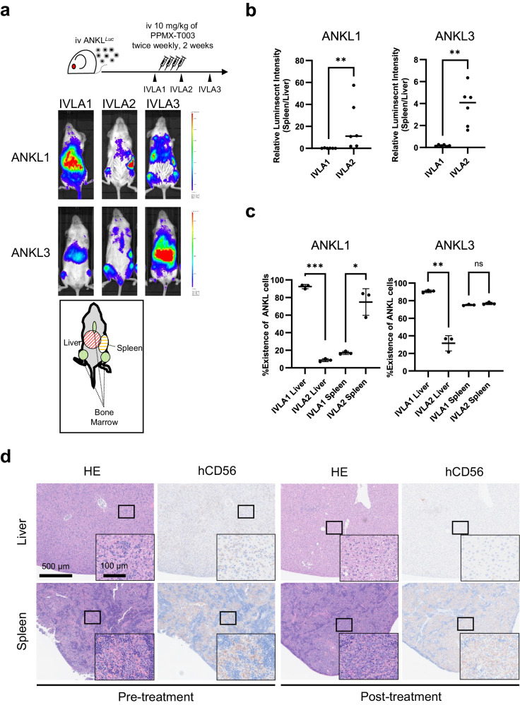
Fig. 1. ANKL cells in the spleen and bone marrow were resistant to treatment with an anti-TfR1 antibody, PPMX-T003.
a In vivo luciferase assay (IVLA) of pre- (IVLA1), early after- (IVLA2), and late after- (IVLA3) treatment of two ANKL-PDXs (ANKL1-PDX and ANKL3-PDX) with PPMX-T003 as previously reported [8]. b Relative luminescent intensities of the spleen compared with the liver of six ANKL1-PDXs and six ANKL3-PDXs at the time of IVLA1 and IVLA2 in (a). Setting of the regions of interest was shown in Supplementary Fig. S1. c Proportion of human CD45-positive cells (i.e., ANKL cells) of liver- and spleen-derived cell suspension of ANKL1- and ANKL3-PDXs at IVLA1 and IVLA2. Hepatocytes were excluded by density gradient centrifugation, and proportions were measured using flow cytometer (see Supplementary Fig. 1b). Three mice per stage per lineage were analyzed. d Hematoxylin and eosin (HE) staining and immunohistochemistry with an anti-human CD56 antibody of the liver and spleen derived from pre- and post-treated ANKL1-PDX. Squares in pictures of low-powered fields indicate the place of high-powered fields.

