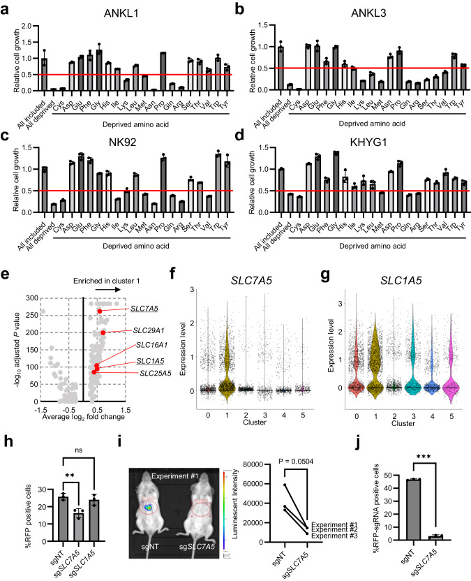
Fig. 4. LAT1-mediated amino acid influx positively regulates ANKL cell proliferation.
a–d In vitro cell proliferation assay of liver-derived ANKL cells and ANKL-derived cell lines cultured in single amino acid-deprived RPMI 1640. Each bar indicates the relative proliferation value to all amino acid-included conditions. The red line indicates a relative proliferation value of 0.5, a cut-off of these assays. Total of three samples per treatment group were analyzed. e Volcano plot of feature genes of cluster 1 of single-cell whole-transcriptome analysis described in Fig. 3, compared with other clusters. Red dots and underbars indicate SLC family genes and genes encoding amino acid transporter, respectively. f–g Violin plots of SLC7A5 (h) and SLC1A5 (i) of single-cell whole-transcriptome analysis described in Fig. 3. h In vitro competitive proliferation assay of transduced Cas9-overexpressed NK92 cells with sgRNA-RFP plasmids by lentiviral vector. Proportions of RFP-positive cells (i.e., target gene-knocked-out cells) were measured using flow cytometry two and eight days after lentiviral transduction. %RFP-positive cells is determined by dividing the number of RFP-positive cells of day 8 by those of day 2. Total of three samples per treatment group were analyzed. i In vivo luciferase assay of ANKL3-PDXs established with Cas9-overexpressed ANKL3 cells transduced with sgNT- or sgSLC7A5-RFP plasmids. Assays were performed seven days after ANKL3 cell inoculation. Luminescent intensities of liver regions were measured. Experiments were independently performed three times, and statistical value was calculated using paired t-test. j Proportions of RFP-positive cells (i.e., target gene-knocked out cells) of liver-derived ANKL3 cells at the timing of in vivo luciferase assay in (h), measured using flow cytometry.

