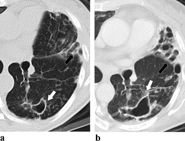
Fig. 2.

Serial changes in cavitating lung lesions in a 53-year-old female with Mycobacterium abscessus complex lung disease (MABC-LD) in the treatment failure group (without microbiological conversion). The follow-up period was 2 years. a Initial CT scans (section thickness, 5 mm, in axial plane) revealed a thin wall cavitation (white arrows) in the left lower lobe with severe bronchiectasis (black arrows) in the lingula left upper lobe. The largest diameter of cavitation is 5.44 cm in the apical left upper lobe (not shown), scoring 3 in cavitation diameter. For cavitation extension, involvement of two segments of the left upper lobe and one segment of the lingula lobe (not shown) earned 2 and 2 points, respectively. Additionally, involvement of all segments of the left lower lobe earned 3 points. This totals 7 points. Therefore, the final cavitation extension score is 3. b Follow-up CT scan (section thickness, 5 mm, in axial plane) at a similar level showed enlargement of the cavity and the bronchiectasis
