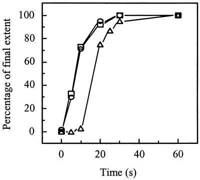
FIG. 8.
Sequence of events after acidification of a SIN-liposome mixture. The kinetics of SIN-liposome binding (squares), E1 trimerization (circles), and fusion (triangles) are shown after acidification to pH 5.75 at 20°C. To compare the kinetics of these processes, the final extents of the relative values of the three parameters were set to 100%. The absolute final extents were 30% for SIN-liposome binding, 56% for E1 trimerization, and 14% for fusion. In each case, SIN (0.5 μM viral phospholipid) was incubated with liposomes (200 μM liposomal phospholipid) consisting of PC/PE/SPM/Chol (molar ratio, 1.0:1.0:1.0:1.5) at pH 5.75 at 20°C. SIN-liposome binding was determined as described in the legend for Fig. 6. E1 trimerization was determined as described in the legend for Fig. 7. Fusion was determined as described in the legend for Fig. 1.

