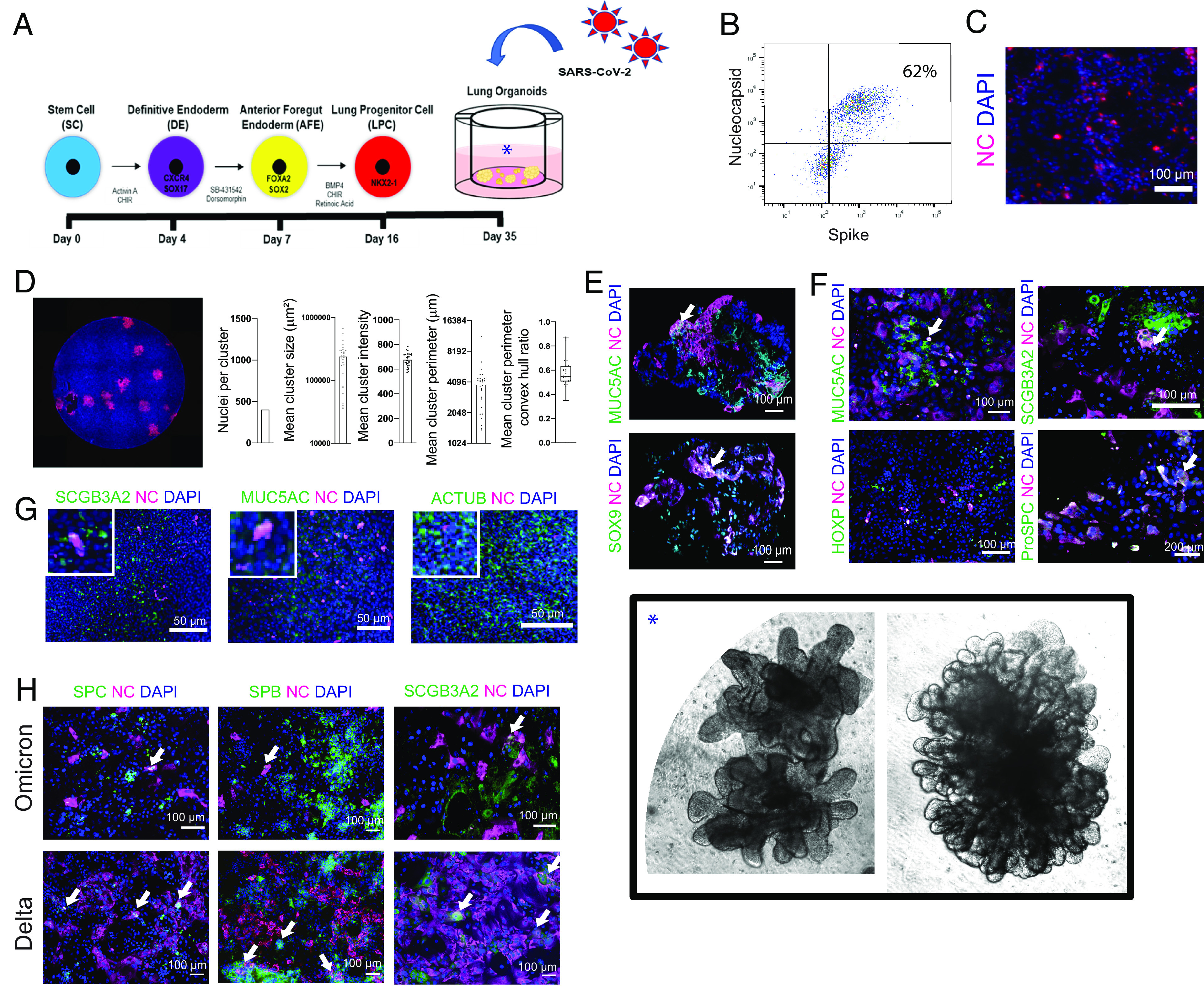
Fig. 1.

hiPSC-derived LOs are susceptible to infection with multiple strains of SARS-CoV-2, validated in primary human lung epithelia. (A) Schematic of steps by which hiPSCs are “instructed” to become 3D LOs that resembles a lung in situ and subsequently infected with SARS-CoV-2. See SI Appendix, Fig. S1, for details. Photomicrographs of actual WLOs are magnified in the Inset [*] in the Lower Right of the montage. (B) Representative flow cytometry of infected 3D PLOs expressing viral proteins Spike and NC confirms that ~62% of the pulmonary epithelial cells were infected. (C) Immunostaining of infected dissociated PLOs 24 hpi for viral NC (cyan) confirms the cytometric findings of viral infection. In all photomicrographs in this figure, a DAPI nuclear stain (blue) is used to visualize all cells in the field. (D) Immunostaining of infected dissociated PLOs overlayed with carboxymethylcellulose to enhance visualization and permit quantification. Cells were fixed, permeabilized, and costained with NC-AF594 antibody (red). A representative well is shown. Images were quantified with a custom-scripted Image J code. Measurements of NC+ nuclei/cluster, cluster size and perimeter, and cluster intensity confirm robust infection. (E) Immunostaining of intact 3D WLOs 36 hpi for coexpression of viral NC (cyan) (indicative of infection) in multiple pulmonary epithelial cell types as identified by cell type-specific immunomarkers (green). Two representative cell types are shown: goblet cells (MUC5AC) and LPCs (SOX9). Note that triple-labeled cells (NC [cyan] plus cell type marker [green] plus DAPI [blue]) typically appear white (an example of which is indicated by an arrow in this and the other Panels). (F) Immunostaining of dissociated 3D PLOs and DLOs with viral NC and cell-type lung epithelial markers: goblet cells (MUC5AC), club cells (SCGB3A2), alveolar type 2 cells (proSPC), and AT1 cells (HOPX). The arrow indicates representative triple-labeled cells. (G) To validate the range of cell types infected in LOs, primary human bronchial epithelial cell (HBEC) ALI cultures were also infected and immunostained 24 hpi. Shown in the coexpression of viral NC (evidence of infection) in a range of the same representative pulmonary epithelial cells shown above: goblet cells, club cells, and ciliated cells (AcTub). Insets are 2.5× original images. (H) Immunostaining of dissociated PLOs and DLOs 24 hpi, comparing SARS-CoV-2 variants Omicron vs. Delta. (Arrows indicate infected cells). The data are representative of at least three independent experiments. All scale bar, 100 µm unless otherwise specified.
