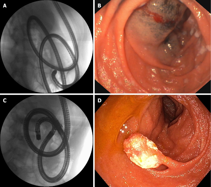
Figure 2.
A 21-year-old female patient with juvenile polyposis associated with Osler–Weber–Rendu disease (hereditary hemorrhagic telangiectasia) and a 10-mm hamartomatous polyp of the jejunum. A: Single balloon enteroscopy was used to reach the medium jejunum; B: A tattoo with submucosal ink was placed at the maximum insertion depth; C: A few months later, the same endoscopist performed motorized spiral enteroscopy, reaching the distal jejunum: D: The polyp could be detected and easily removed with standard polypectomy. Note the different looping of the scope in the fluoroscopic view.

