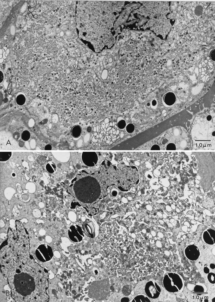
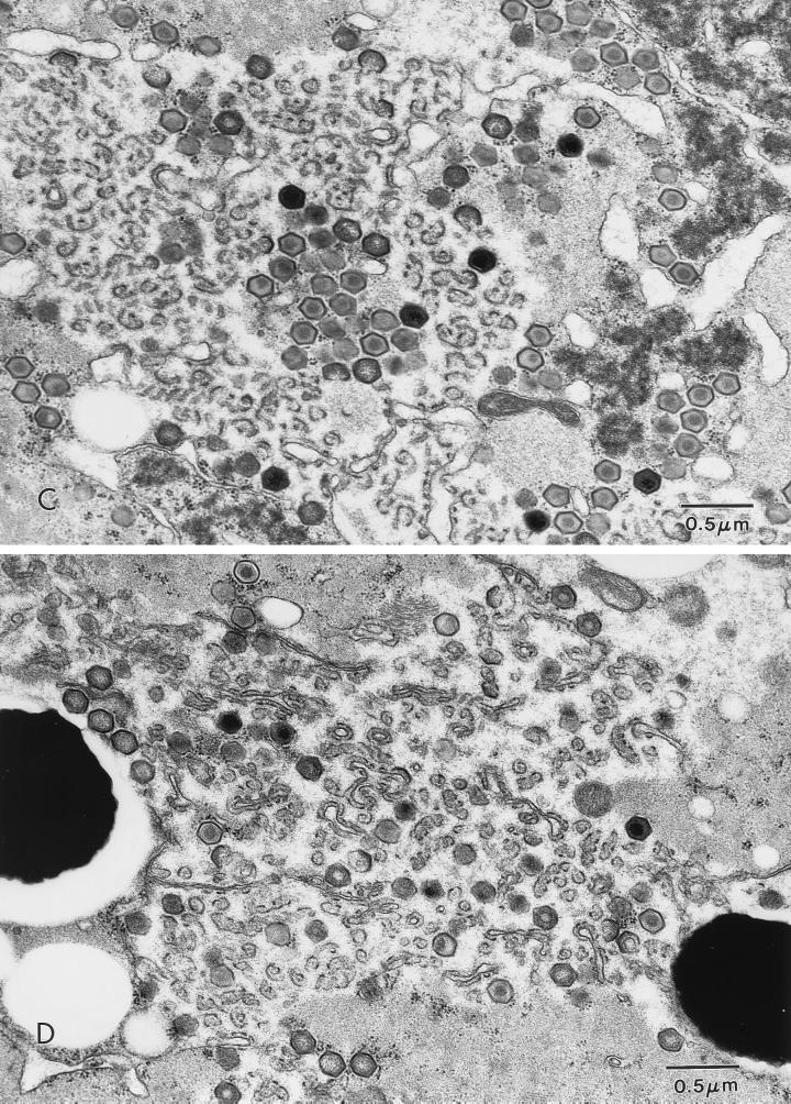
FIG. 3.
ASFV in an O. porcinus porcinus midgut. Ticks were exposed by membrane feeding. The inoculum contained Pr4 (A and C) or MAL (B and D). Analysis was performed 6 days p.i. (A) Virus factory (VF) in a midgut epithelial cell from a Pr4-exposed tick. (B) Virus factory in a midgut epithelial cell from an MAL-exposed tick. (C) Higher magnification of a virus factory from a Pr4-exposed tick. (D) Higher magnification of a virus factory from an MAL-exposed tick. N, nucleus; L, midgut lumen; NF, nucleus free in the midgut lumen.


