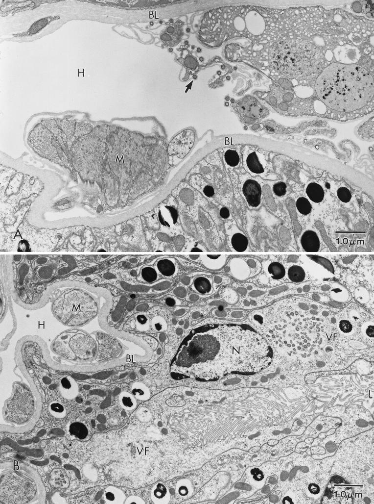
FIG. 5.
ASFV in an O. porcinus porcinus midgut. Analysis was performed 14 weeks after intrahemocoelic inoculation. (A) Mature virions (arrow) budding from a connective tissue cell adjacent to the basal lamina (BL) of the midgut in an MAL-injected tick. (B) Virus factory (VF) in a midgut epithelial cell from a Pr4-injected tick. H, hemocoel; L, lumen; M, muscle; N, nucleus.

