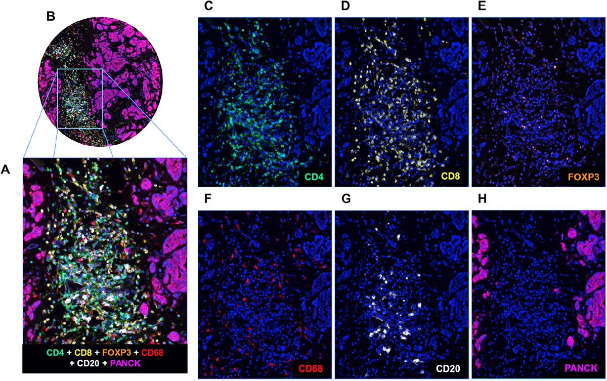
Figure 1.

mIF staining of a panel of five immune markers plus one epithelial marker. (A and B) A breast cancer TMA core showing composite staining of each of the six markers in the mIF panel (CD4, CD8, F0XP3, CD68, CD68, and PanCK) together with DAPI. (C–H) Individual images of CD4 (green), CD8 (yellow), F0XP3 (orange), CD68 (red), CD20 (white), and PanCK (purple) with DAPI counterstain.
