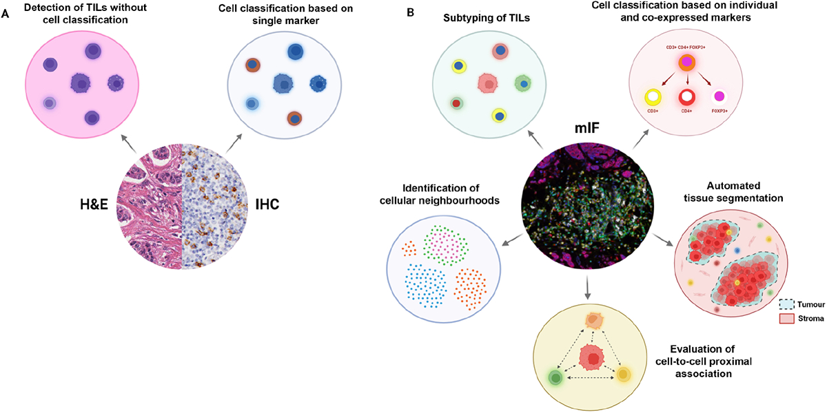
Figure 3.

TIL profiling with H&E versus conventional chromogenic IHC versus mIF. (A) H&E staining enables the measurement of total TILs in tissue, whereas, with conventional IHC, a specific immune population can be profiled based on a single protein marker. (B) mIF staining allows the total TIL population to be subtyped based on multiple markers. It is also possible to further characterise cells based on marker colocalisation. With an epithelial or tumour differentiation marker, it is possible to automate tumour-stroma segmentation with image analysis software. Furthermore, multiplex images can be used to map spatial distributions of different cell phenotypes and examine their proximal associations, and further identify distinct cellular neighbourhoods. Created with BioRender.com.
