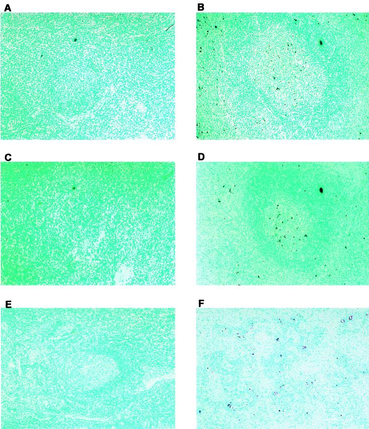
FIG. 2.
In situ detection of cell death in lymph nodes from mock-infected and BHV-1-infected calves. Thin sections were prepared as described for Fig. 1 from cervical lymph nodes from a mock-infected (A) and a BHV-1-infected calf (B), retropharyngeal lymph nodes from a mock-infected (C) and a BHV-1 infected calf (D), and inguinal lymph nodes from a mock-infected (E) and a BHV-1 infected calf (F). All samples are at a magnification of ×100. Methyl green was used to counterstain. Dark purple color indicates the TUNEL+ cells.

