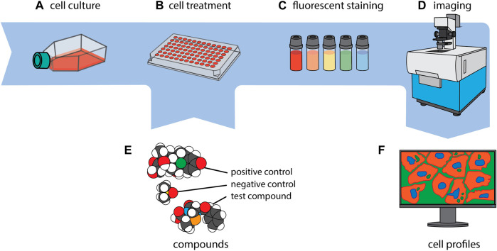
FIGURE 1.
CP in a nutshell. Cells of interest are first cultivated (A) and then seeded to, typically, a 384-well plate (B). Every well is exposed to a single compound or genetic perturbation (E) and incubated for a period ranging from 24 to 48 h, after which every well is uniformly stained with a collection of fluorescent dyes (C). Subsequently, imaging of the cells is carried out (D), and a phenotypic profile is generated for each experimental condition (F). The images of the molecules in section (E.) were created using CineMol (Meijer et al., 2024).

