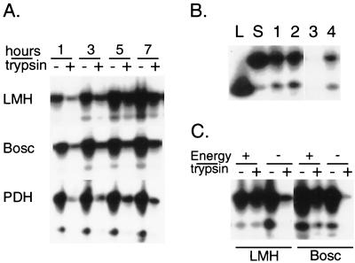
FIG. 6.
Internalization of DHBV particles into DCPD-reconstituted cell lines. (A) Total and trypsin-resistant fractions of DHBV DNA at different time points of virus incubation. DCPD-reconstituted Bosc and LMH cells as well as freshly plated PDH were incubated at 37°C with a 1:10 dilution of 60 μl viremic duck serum for 1 to 7 h. Half of the cell pellet was pretreated with trypsin before DNA extraction. (B) DHBV entry at 4°C and at 37°C. Two wells of DCPD-transfected LMH cells were incubated with a 1:10 dilution of viremic duck serum at 4°C for 2 h. After washing, cells from one well (lanes 1 and 3) were removed, and half of the cell pellet (lane 3) was treated with trypsin. Cells in the other well (lanes 2 and 4) were further incubated at 37°C for 2 h and half of the cell pellet (lane 4) was treated with trypsin. Lanes L and S are as in Fig. 4. (C) Energy depletion inhibits DHBV entry. DCPD-reconstituted LMH cells were preincubated for 1 h with 600 μl of medium in the presence (energy −) or absence (energy +) of sodium azide (0.1%) and 2-deoxy-d-glucose (50 mM) and incubated for 3 h following the addition of 60 μl of viremic duck serum. The DCPD-transfected Bosc cells were preincubated with energy depleting agents for 3 h and incubated for 12 h with DHBV. Both total and trypsin-resistant fractions of DHBV DNA were measured.

