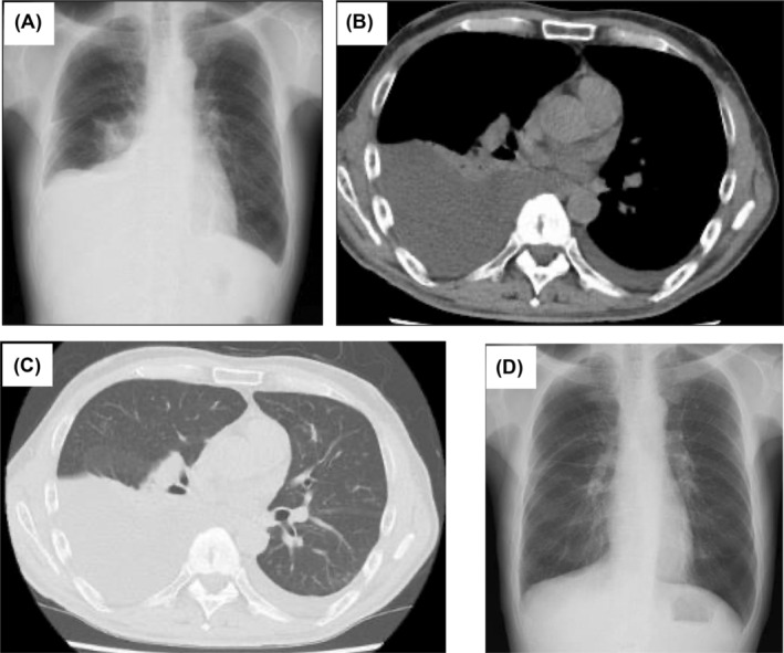
FIGURE 1.

(A) Chest radiograph on admission showing right‐dominant bilateral pleural effusions. (B) and (C) Chest computed tomography scan showing right‐dominant bilateral pleural effusions without any parenchymal lesions. (D) The pleural effusion was improved on a chest radiograph after 1 month of corticosteroid therapy (35 mg/day oral prednisolone).
