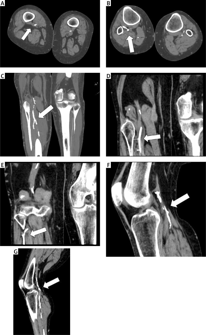
Figure 1.
Computed tomography images revealing arterial occlusion of the lower limb by foreign bodies (arrows). A – Transverse images of the distal thigh showing the foreign body in the superficial femoral artery. B – Transverse images showing the foreign body at the knee level. C–E – Coronary reconstructions. F, G – Sagittal reconstructions

