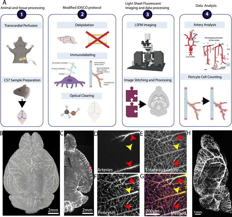
Fig. 4. Tissue clearing and 3D immunolabeling with LSFM imaging to examine different vascular compartments and mural cells in the same brain.
A The steps of brain clearing, whole brain immunolabeling, and light sheet fluorescent microscopy (LSFM) pipeline are outlined in order. 1. Brain sample collection with transcardial perfusion. 2. Modified iDISCO protocol including delipidation, immunolabeling for arteries, whole vasculature, and pericytes, and optical clearing. 3. LSFM imaging and data processing to visualize cleared brains at cellular resolution. 4. Data analysis such as arteriole geometry analysis and pericyte counting. B 3D reconstruction of a brain with artery staining by LSFM imaging, scale bar 2 mm. C Max projection of the 500 μm thick z stack of the artery staining, scale bar 2 mm. D–G Zoom-in images of the red box area from (C), scale bars 200 μm. D artery staining in the green channel, E lectin based total vasculature staining in the red channel, F pericyte staining in the far-red channel, G a merged image of pseudo-colored arteries (blue), total vasculature (green), and pericyte (red). H Maximum projection of the artery channel in a brain hemisphere, scale bar 1 mm.

