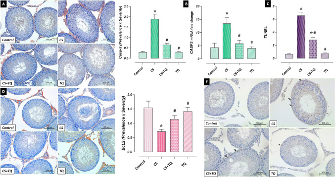
Fig. 8.
Effect of CS and/or TQ applications on apoptotic markers in testicular tissues: A; Casp3 immunoreactivity microphotographs and graph, B; Casp3 mRNA expression, C; TUNEL graph, D; BcL2 immunoreactivity microphotographs and graph, E; TUNEL microphotographs. Levels of apoptotic markers in testicular tissues of control and TQ groups were similar. It was determined that BcL2 immunoreactivity decreased in the CS group compared to the control group, while Casp3 levels and TUNEL-positive apoptotic cells increased. In the CS + TQ group, it was observed that BcL2 immunoreactivity increased compared to the CS group, while Casp3 levels and TUNEL-positive apoptotic cells decreased. *; compared to the control group (p < 0.05), #; Compared to CS group (p < 0.05). A; Casp3 immunohistochemical staining, D; BcL2 immunohistochemical staining, E; TUNEL staining, scale bar; 100 μm. CS; Cisplatin, TQ; Thymoquinone

