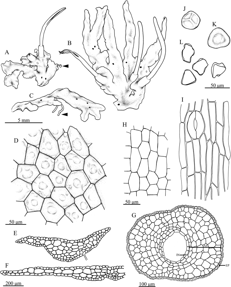
Figure 3.
Phaeocerosperpusillusvar.scabrellusA gametophyte forming half-rosettes with sporophyte (arrow indicate tuber) B ensiform thalli and sporophytes C gametophyte showing ventral tuber (arrow) D dorsal epidermal cells of thallus E, F cross sections of thalli G cross section of sporangium (AS = assimilative tissue, EP = epidermal cell of capsule, IN = inner most sporangium wall) H inner most cells of sporangium wall I epidermal cells of capsule with stoma J proximal view of spore K distal view of spore L pseudoelaters. All from holotype and drawings by O. Suwanmala. (All drawing from S. Chantanaorrapint & O. Suwanmala 4116).

