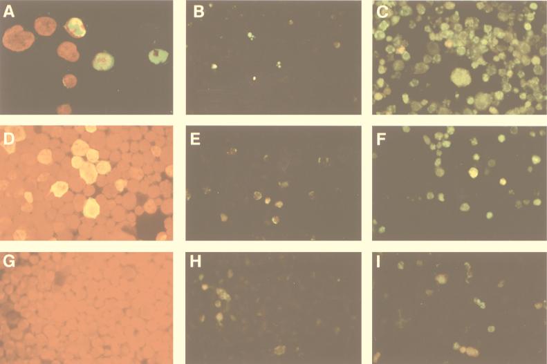
FIG. 1.
(A) Expression of NS-1 protein in B19-infected CD36 cells. Magnification, ×400. The cells were fixed and incubated with a polyclonal rabbit anti-NS-1 antibody, and expression was revealed with monoclonal fluorescein isothiocyanate (FITC)-conjugated anti-rabbit Ig and then DAPI (4′,6-diamidino-2-phenylindole). Forty-eight hours after infection, NS-1 protein (green) was detected by confocal microscope analysis in the nucleus (red) and the cytoplasm in approximately 50% of the cells. (B) Apoptosis in uninfected CD36 cells. Magnification, ×250. Fragmented DNA of fixed cells was labeled by adding fluorescein dUTP, using the TUNEL assay. (C) Apoptosis in B19-infected CD36 cells 72 h after infection. (D) NS-1 protein expression in UT7-NS cells 48 h after its induction by DEX. The cells were fixed and incubated with a polyclonal rabbit anti-NS-1 antibody. NS-1 was revealed with monoclonal FITC-conjugated anti-rabbit Ig by immunofluorescence microscopy. (E) Apoptosis in uninduced UT7-NS cells. (F) Apoptosis in DEX-induced UT7-NS cells at 72 h. (G) UT7-pGRE cells incubated with a polyclonal rabbit anti-NS-1 antibody and a monoclonal FITC-conjugated anti-rabbit Ig antibody and then revealed by immunofluorescence microscopy. (H) Apoptosis in uninduced UT7-pGRE cells. (I) Apoptosis in DEX-induced UT7-pGRE cells.

