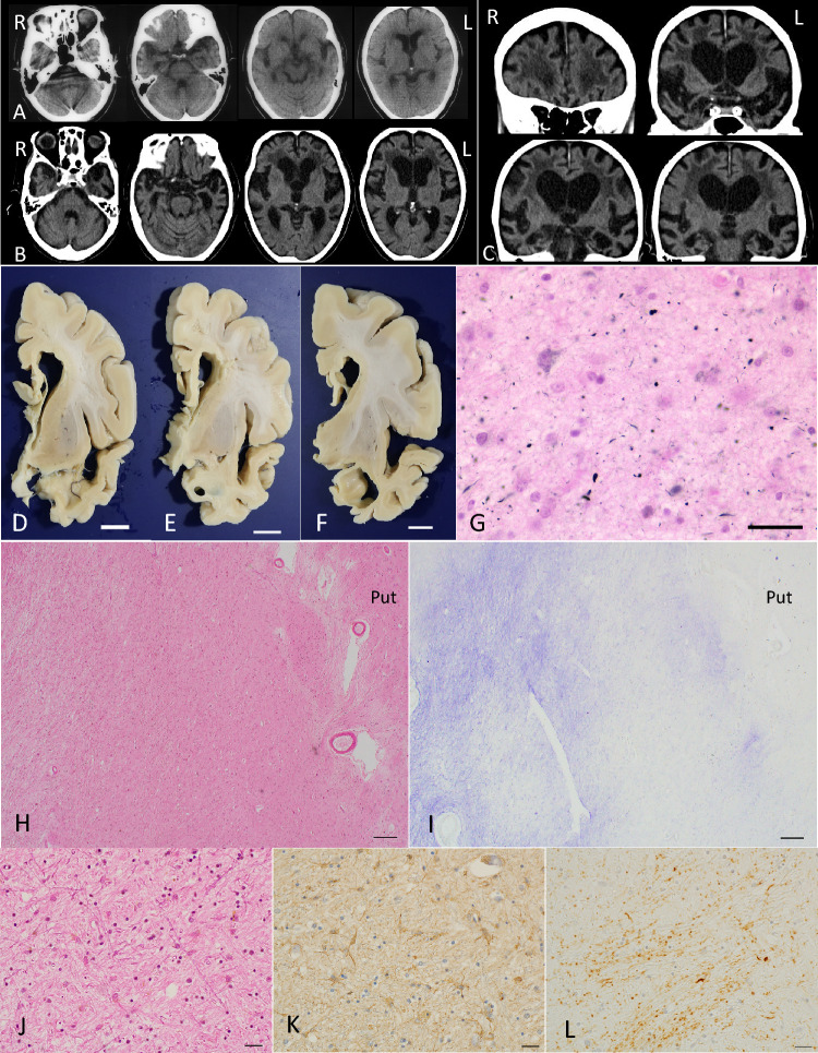
Fig. 1.
Radiological and pathological findings in case 1. A Axial CT images at the age of 74 years. Mild atrophy was noted in the amygdala, but the neocortex was almost completely spared. Bilateral Sylvian fissures were slightly dilated. B, C Axial (B) and coronal (C) CT images at age 86. Atrophy in the amygdala became severe. Atrophy in the neocortex was diffuse and symmetric but was slightly accentuated in the frontotemporal lobes. D–F Macroscopic findings of coronal sections of the left hemisphere. Scale bars = 1 cm. G AGs in the amygdala. Gallyas method. Scale bar = 30 μm. H Severe neuronal loss with gliosis in the globus pallidus. The degeneration is more evident in the internal segment (especially in the dorsal portion) than in the external segment in the site. Put: the putamen. H&E stain. Scale bar = 200 μm. I The same region shown in (H). Fibrillary gliosis is more evident in the internal segment rather than the external segment in the globus pallidus. Holzer stain. Scale bar = 200 μm. J–L Severe neuronal loss with gliosis in the globus pallidus revealed by H&E stain (J) and GFAP immunohistochemistry (K). Tau accumulation in the same region (L). AT8 immunohistochemistry. All scale bars = 20 μm. A–C Reprinted with permission from reference [66]

