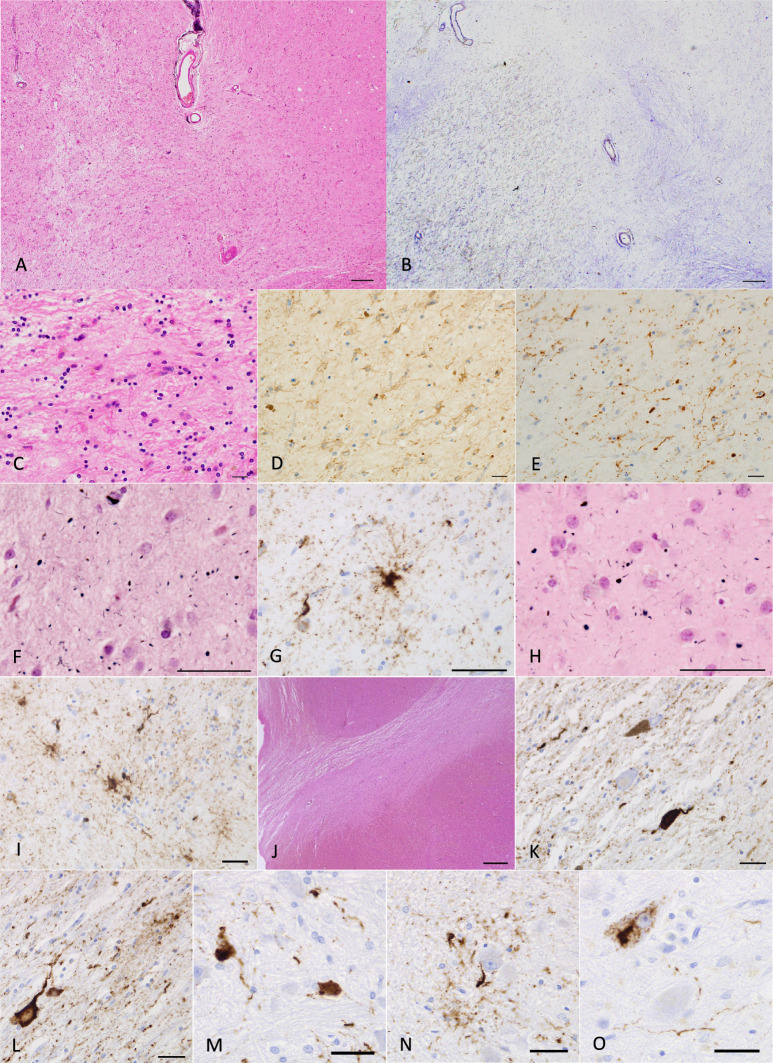
Fig. 4.
Pathological findings in case 2. A Severe neuronal loss with gliosis in the globus pallidus. The degeneration is more evident in the internal segment (left side) rather than external segment (right side) in the site. Especially, tissue rarefaction is severe in the dorsal portion of the internal segment. H&E stain. Scale bar = 200 μm. B Fibrillary gliosis is more evident in the internal segment than in the external segment in the globus pallidus. Holzer stain. Scale bar = 200 μm. C, D Severe gliosis in the globus pallidus demonstrated by H&E stain and GFAP immunohistochemistry. Scale bars = 20 μm. E AT8-positive threads and dot-like lesions in the globus pallidus. Scale bar = 20 μm. F AGs in the caudate nucleus. Gallyas method. Scale bar = 50 μm. G AT8-positive GFAs and a coiled body in the caudate nucleus. Scale bar = 50 μm. H Argyrophilic grains in the putamen. Gallyas method. Scale bar = 50 μm. I AT8-positive GFAs in the putamen. Scale bar = 50 μm. J Severe neuronal loss with gliosis in the substantia nigra. H&E stain. Scale bar = 600 μm. K AT8-positive NFT, threads, and AGs in the substantia nigra. Scale bar = 30 μm. L AT8-positive NFT, GFA, and threads in the subthalamic nucleus. Scale bar = 30 μm. M AT8-positive NFTs and threads in the pontine nucleus. Scale bar = 30 μm. N AT8-positive GFA and coiled body in the inferior olivary nucleus. Scale bar = 30 μm. O AT8-positive NFT and threads in the dentate nucleus in the cerebellum. Scale bar = 30 μm

