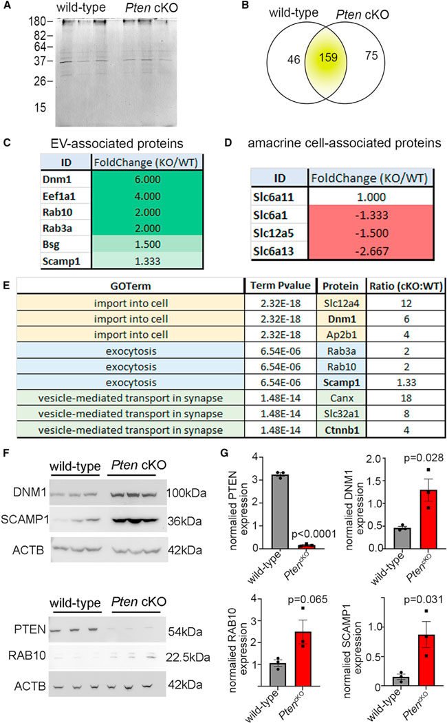
Figure 5. The proteome of the retinal vesiculome reveals an enrichment of endocytic trafficking proteins in PtencKO retinal cells.

(A) Silver-stained SDS-PAGE gel with EV lysates purified from P14 wild-type and PtencKO retinas.
(B) LC-MS/MS analyses of EV proteins isolated from P14 wild-type and PtencKO retinas, showing the numbers of proteins enriched in wild-type and PtencKO samples.
(C and D) EV-associated proteins (C) and amacrine cell markers (D), showing fold change, comparing PtencKO/wild-type retinas.
(E) Biological process GO terms associated with enriched proteins in PtencKO retinal EVs.
(F and G) Western blots for PTEN, RAB10, DNM1, and SCAMP1 in P14 wild-type and PtencKO retinas. Plots show mean densitometry values normalized to ACTB ± SEM. N = 3 biological replicates/genotype.
The p values were calculated with unpaired t test. See also Figure S6.
