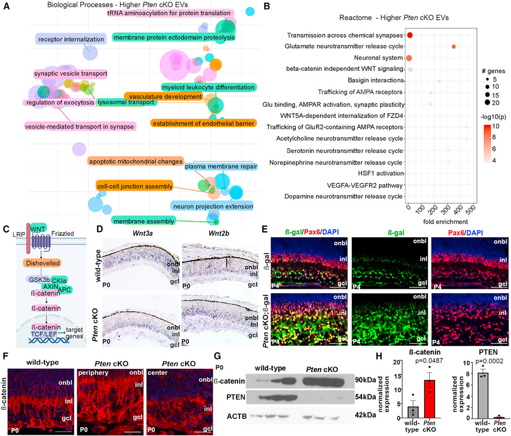
Figure 6. Canonical Wnt signaling is negatively regulated by Pten and active in migrating amacrine cells.

(A) Biological processes enriched in PtencKO retinal EVs.
(B) Reactome pathway analysis of enriched proteins in PtencKO retinal EVs. The size of the circle represents the number of genes enriched in the pathway; the color of the circle represents the log10 p value.
(C) Schematic of the canonical Wnt signaling pathway.
(D) Expression of Wnt3a and Wnt2b in P0 wild-type and PtencKO retinas using RNA in situ hybridization.
(E) Expression of β-galactosidase (β-gal) and Pax6 in P4 lef/tcf::lacz and PtencKO; lef/tcf::lacz retinas.
(F) Expression of β-catenin in P10 wild-type and PtencKO retinas, including in the peripheral (region of Pten deletion) and central (Pten expression is retained) retina.
(G and H) Western blots of β-catenin and PTEN in P0 wild-type and PtencKO retinas.
Plots show mean densitometry values normalized to ACTB ± SEM. N = 3 biological replicates/genotype. The p values were calculated with an unpaired t test. Scale bars: 100 μm (E and F).
