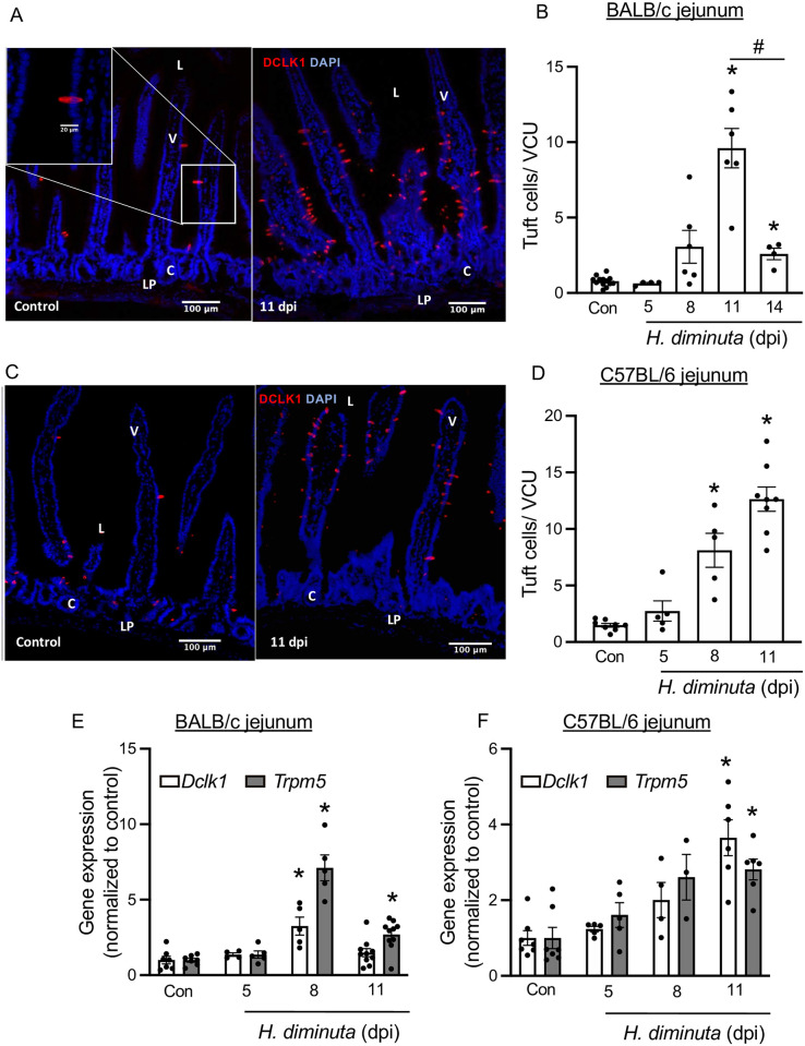
Fig 2. H. diminuta-infection induces murine small intestinal tuft cell hyperplasia.
Male BALB/c and C57BL/6 mice were infected with 5 cysticercoids of H. diminuta and assessed at days post-infection (dpi). (A, C) Representative images of mid-jejunal cryosections (10 μm) from control and infected BALB/c and C57BL/6 mice (11 dpi) immunostained for DCLK1 (red) and counterstained with DAPI (blue). “L”–lumen, “V”- villus,” C”- crypt, “LP”- lamina propria and “S”- serosa on the image. DCLK1+ cells were enumerated per villus crypt unit (VCU) and averaged over 20 respective units per mouse. (E, F) Relative mRNA expression (compared to the 18S rRNA housekeeping gene) in mid-jejunal segments from BALB/c and C57BL/6 mice respectively, was assessed by real-time PCR. Data are mean ± SEM values, n = 4-9/group, pooled from 2–3 experiments, * p<0.05 compared to the control (Con) group, analysed by (B, D) Browns Forsythe and Welch’s ANOVA and Dunnett’s test or (E, F) Kruskal Wallis test and Dunn’s test for multiple comparisons; # p<0.05 comparing tuft cell counts at 14 dpi vs at 11 dpi analysed by Welch’s t test (A).

