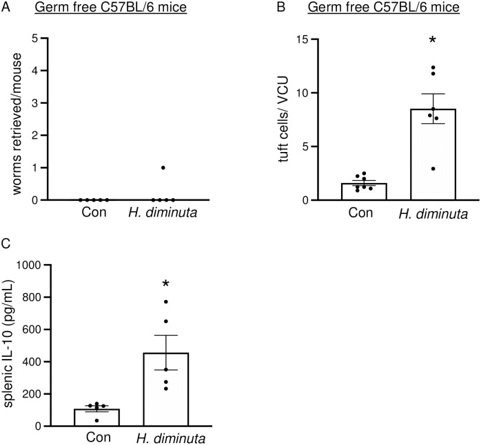
Fig 4. Tuft cell hyperplasia in response to H. diminuta does not depend on the microbiome.
Male germ free C57BL/6 mice were infected with 10 antibiotic-treated cysticercoids of H. diminuta and assessed at 11 days post-infection (dpi). (A) Murine small intestines were flushed with ice cold PBS at necropsy to retrieve and enumerate worms. (B) Mid-jejunal sections were immunostained with anti-DCLK1 antibody and counterstained with DAPI. DCLK1+ cells were enumerated per villus crypt-unit (VCU) averaged over at least 3 random VCUs/mouse. (C) IL-10 ELISA was performed on supernatants from splenic cells (5x10⁶/mL) stimulated with concanavalin A (2 μg/mL, 48h). Data are mean ± SEM values, n = 5-8/group, pooled from 2 experiments, * p<0.05 compared to control (Con) mice and analysed by Welch’s unpaired t test.

