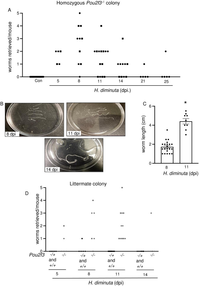
Fig 5. Tuft cell deficiency delays expulsion of H. diminuta from the murine intestine.
Male (●) and female (◼) Pou2f3-/- (homozygous colony) (A-C), and Pou2f3+/+, +/-, -/- littermate mice (D) were infected with 5 cysticercoids of H. diminuta and assessed at days post-infection (dpi). (A, B) Small intestines were flushed with ice cold PBS to retrieve and enumerate worms. (C) H. diminuta worms from Pou2f3-/- mice were significantly longer at 11 dpi. (D) Unlike their infected wild type littermates that show no detectable worms, Pou2f3-/- mice still harbor H. diminuta worms at 11 dpi. Data in (C) are mean ± SEM, n = 9-21/group, pooled from 2–3 experiments, *p<0.05 analysed by Welch’s t test.

