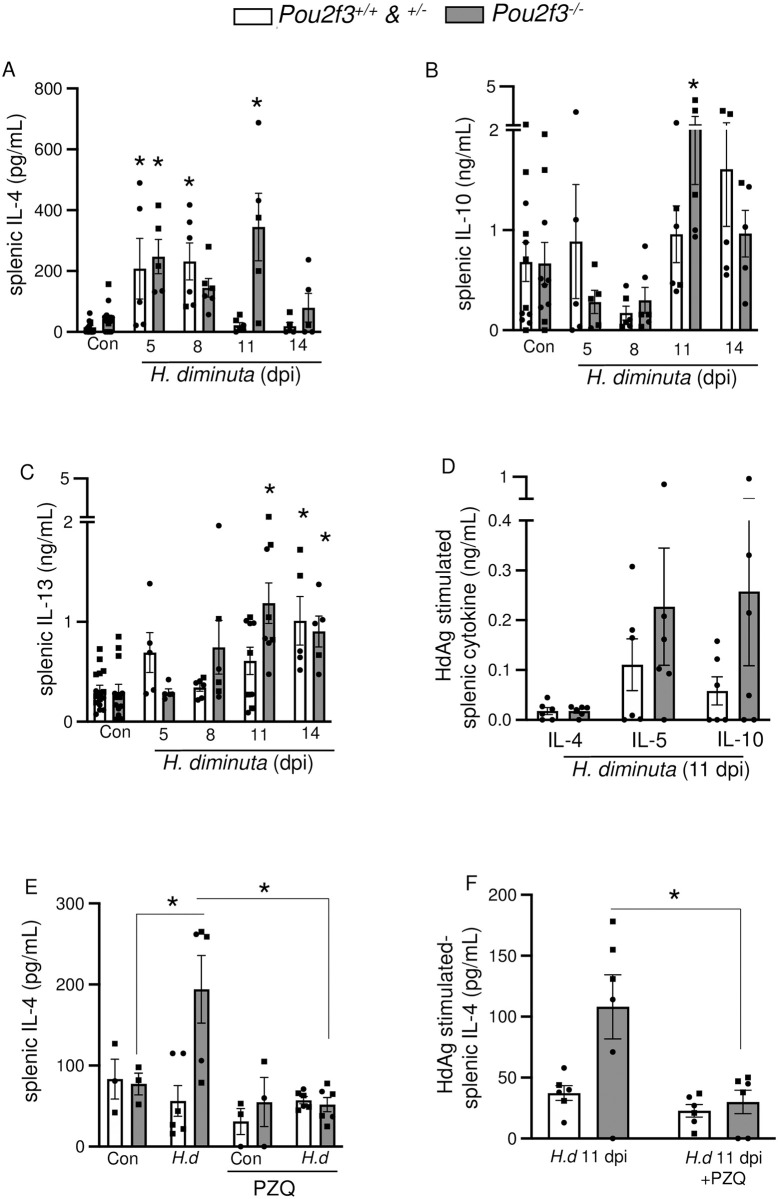
Fig 7. Tuft cell-deficient mice display systemic Th2 immune response following infection with H. diminuta.
Male (●) and female (◼) Pou2f3+/+, +/-, -/- mice were infected with 5 cysticercoids of H. diminuta (H.d) and assessed at days post-infection (dpi). In (E, F), mice were treated with praziquantel (PZQ; 1 mg/mouse by oral gavage) at 8 dpi prior to necropsy on 11 dpi. (A, B, C, E) Cytokine ELISAs for IL-4, IL-10, and IL-13 were performed on supernatants from splenic cells (5x10⁶) stimulated with concanavalin A (2 μg/mL, 48h) or (D, F) a PBS-soluble crude extract of adult H. diminuta (HdAg, 200 μg/mL, 96h). Data are mean ± SEM, n = 3-6/group, pooled from 1–3 experiments, * p<0.05 compared to control (Con) uninfected mice, or as indicated on the graph, analysed by (A, B, C) Two-Way ANOVA and Dunnet’s/ Tukey’s post-test or (D) multiple Welch’s t tests.

