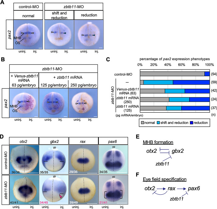
Fig 5. Effects of zbtb11 knockdown on anteroposterior patterning of the neuroectoderm and eye field formation.
zbtb11-MO or control-MO was injected into the animal pole region of one dorsal blastomere at the 4-cell stage. (A-C) WISH analysis for pax2 expression was performed at the late neurula stage (st. 19–23). (A) Representative images of pax2 expression in control-MO- or zbtb11-MO-injected embryos. The mode of pax2 expression was categorized: ‘normal’, ‘shift and reduction’, and ‘reduction’. (B,C) mRNA rescue experiments. Venus-zbtb11 mRNA (63 pg/embryo) or zbtb11 mRNA (125 or 250 pg/embryo) was co-injected with nβ-gal mRNA into the dorsoanimal blastomere on the same side as the MO-injected side at the 8-cell stage. (B) Representative images of pax2 expression in zbtb11 morphants injected with mRNAs. Red-gal stained cells indicate Venus-zbtb11 or zbtb11 expressing cells. White arrowheads, pax2 expression at the MHB; blue arrowheads, reduction of pax2 at the MHB on the injected side (A,B). (C) Venus-zbtb11 or zbtb11 mRNA partially rescues the abberant pax2 expression in zbtb11 morphants. Amounts of injected Venus-zbtb11 mRNA or zbtb11 mRNA are as indicated. Biologically independent experiments were repeated twice (A-C). (D) WISH analysis for otx2, gbx2, rax and pax6 was performed at the early neurula stage (st. 13–14). Biologically independent experiments were repeated 4 to 6 times. Fractions indicate the numbers of the embryos presenting the phenotype per scored embryos (numbers in white or black, minor effects on gene expression; magenta, expanded expression). Dashed line, the midline of the embryo; solid line, the anteriormost position of gbx2 expression; magenta arrowhead, anterior expansion of gbx2; bracket, the size of the eye field expressing pax6. Anterior view with the dorsal side up (pax2, otx2, rax), and dorsal view with the posterior side up (gbx2, pax6). MHB, the midbrain and hindbrain boundary; OS, optic stalk; yp, yolk plug. inj., MO-injected side; uninj., uninjected side. n, the total number of each sample (C). Scale bars: 100 μm (A,B), 500 μm (D). Amounts of injected MOs (pmol/embryo): 0.5. (E,F) Schematic models of gene interactions in MHB formation and eye-field specification. Mutual repression between otx2 and gbx2 (E) and the gene cascade of otx2, rax, and pax6 (F) have well been documented (see the text). zbtb11-MO experiments suggest that Zbtb11 represses gbx2 expression anterior to the MHB and represses pax6 but not rax in the eye field. Arrow, activation; T mark, inhibition.

