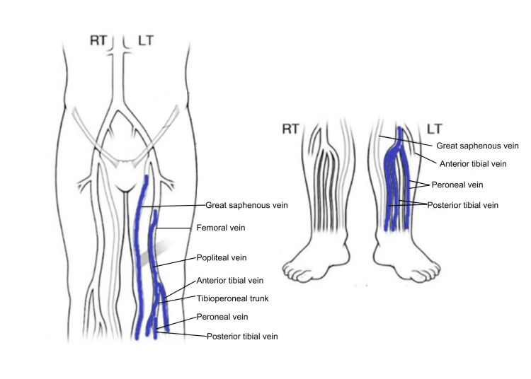
Figure 1. Ultrasound Doppler study showing multiple deep vein thrombosis. There was a partially occlusive thrombus in the femoral vein mid thigh and fully occlusive thrombus in the distal femoral vein to the popliteal vein. There was an occlusive thrombus in the calf vein.
Image reproduced from radiology images obtained on a deep vein thrombus (DVT) study. Mayo Clinic is the copyright holder of the DVT mapping image.

