
Figure 3. Loss of GPR52 leads to altered breast cancer cell organization in Matrigel.
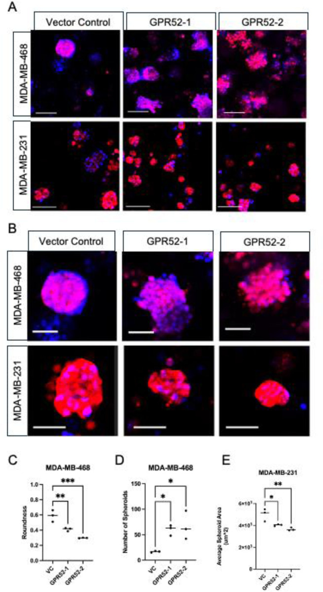
Cells were cultured in Matrigel for 10 days under complete media conditions. (A-B) cytoplasmic tdTomato (red) and 1:1000 nuclear Hoechst 33342 stain (blue) allow visualization of MDA-MB-468 (top row) and MDA-MB-231 (bottom row) WT and GPR52 KO spheroids. (A) Scalebar=100 μm, (B) Scalebar=50 μm. C-E) The roundness, number, and average area of spheroids was determined based on the tdTomato signal. VC=vector control. n=3, One-way ANOVA, P-value<0.05; *P <0.05, **P <0.005, ***P<0.0005, ns=not significant. Line=median.