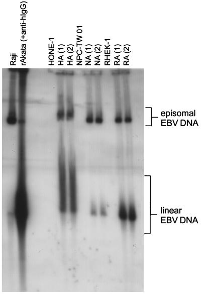
FIG. 4.
Configuration of EBV DNA in virus-infected epithelial cells. Gardella gel analysis was carried out to detect episomal and linear EBV DNA in the cells. Raji cells were used as controls for episomal EBV DNA, and activated rAkata BL cells were used to show linear EBV DNA. HA, NA, and RA cells, as well as the uninfected parental cells, were examined. Two pools from two independent infection experiments for each cell line (numbered above the lanes) were used. Gardella gel analysis was carried out as described previously (8). Briefly, 106 intact cells were loaded into wells surrounded with 2% sodium dodecyl sulfate plus 1 mg of proteinase K per ml in a 0.8% agarose gel and electrophoresed at 4°C for 40 h. DNA in the gel was transferred onto a Hybond-N membrane (Amersham, Little Chalfont, Buckinghamshire, United Kingdom). The blot was hybridized with the random-primed, 32P-labeled EBV BamHI W fragments.

