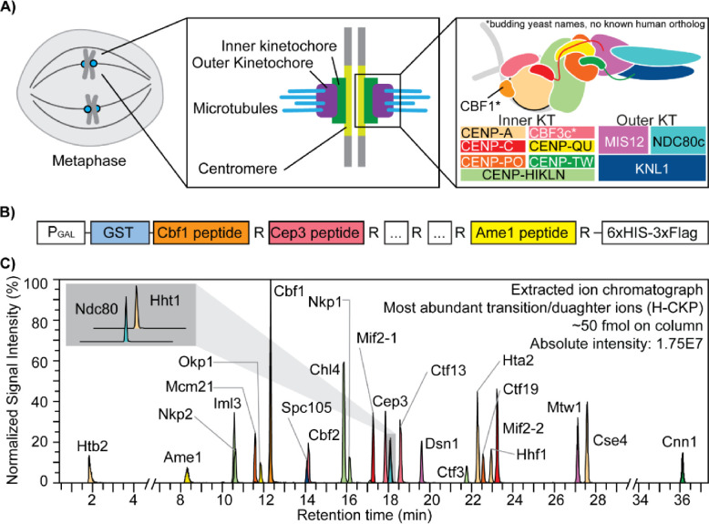
Figure 1. The kinetochore and the concatenated kinetochore protein (CKP) construct.
A) Schematics of yeast kinetochore organization: Yeast kinetochores assemble on the point centromere at each chromosome to carry out mitotic chromosome segregation. The kinetochore can be divided into inner and outer sub-complexes, where the inner kinetochore contacts the centromeric chromatin, and the outer kinetochore forms an association with the spindle microtubules. Both inner and outer kinetochores are composed of multiple protein complexes, as illustrated and annotated in the right-most panel. B) A gene block is constructed to express and purify the concatenated kinetochore protein (CKP): 25 tryptic peptides with a C-terminal arginine were selected from DDA-MS results and incorporated into the CKP. The expression of the protein is controlled by a galactose-inducible promoter (PGAL) in yeast. C) Extracted ion chromatograph of the most abundant daughter ions of H-CKP peptides with retention time in minutes. The y-axis indicates the signal intensities of each ion expressed as a percentage of the signal from the ion with the highest signal intensity (Cbf1 peptide). The total amount of H-CKP injected and the absolute signal intensity for the Cbf1 peptide’s most abundant product ion are labeled on the top right of the plot.

