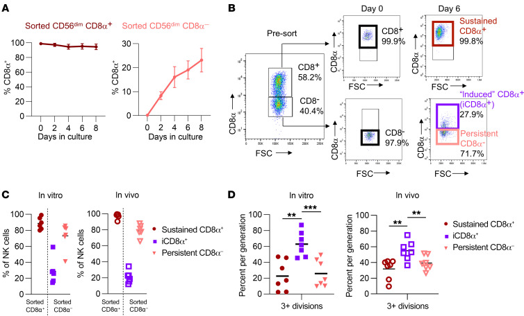
Figure 4. IL-15 modulates CD8α expression.
(A) CD8α+/–CD56dim NK cells were sorted and cultured in 5 ng/mL IL-15 for up to 8 days. Plots show the percentage of NK cells positive for CD8α expression on cells originally sorted as CD8α+ or CD8α– cells. n = 2–3 donors and 2 independent experiments. (B) Gating strategy for identification of induced CD8α+ versus sustained CD8α+ and persistent CD8α– NK cells. Sorted CD8α+ NK cells that remained CD8α+ were defined as sustained CD8α+ cells. Sorted CD8α– NK cells that upregulated CD8α during culturing were defined as induced CD8α+ cells. Sorted CD8α– NK cells that remained CD8α– during culturing were defined as persistent CD8α– cells. FSC, forward scatter. (C and D) CD8α+/–CD56dim NK cells were sorted and cultured in 1 ng/mL IL-15 in vitro or injected into NSG mice supported with i.p. rhIL-15 3 times/week. Data are shown as the percentage of NK cells positive for CD8α expression after 9 days. n = 8 donors and 4 independent experiments. (D) Percentage of NK cells that underwent 3 or more divisions within the indicated subsets in vitro or in vivo in NSG mice 9 days after sorting. n = 6–9 donors and 4 independent experiments. Data represent the mean ± SEM. **P < 0.01 and ***P < 0.001, by 2-way ANOVA with Holm-Šídák correction for multiple comparisons.

