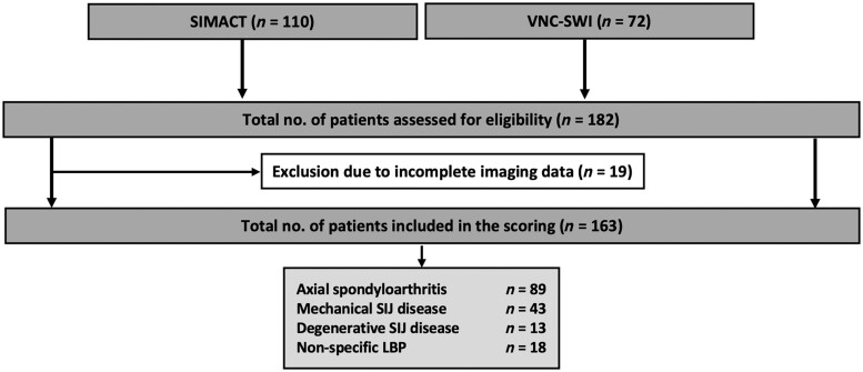
Figure 2.
Flowchart of study inclusion. CT: computed tomography; LBP: low back pain; MRI: magnetic resonance imaging; RG: reader group (based on years of experience in musculoskeletal imaging); RG1: inexperienced reader group; RG2: semi-experienced reader group; RG3: experienced reader group; SIMACT: Sacroiliac Joint Magnetic Resonance Imaging and Computed Tomography Study; VNC-SWI: Virtual Non-Calcium—Susceptibility Weighted Imaging study; XR: X-ray

