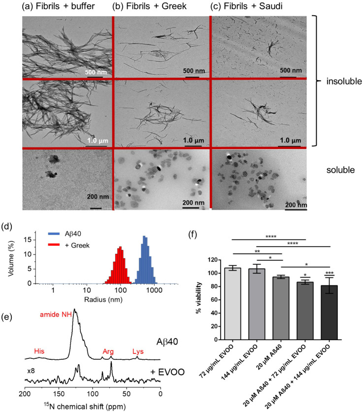
Figure 6.
Negative-stain TEM images of preformed Aβ40 fibrils (20 μM monomer equivalent) incubated for 24 h with either buffer or with 20 μg/mL (≅ 50 μM) EVOO extract. The TEM images show the soluble and insoluble fractions obtained after centrifugation. (a) Aβ40 incubated with buffer. (b) Aβ40 in the presence of Greek EVOO extract. (c) Aβ40 in the presence of Saudi EVOO extract. Two different magnifications and views are shown. (d) DLS data for Aβ40 fibrils alone and following the addition of 740 μg/mL Greek EVOO. (e) 15N CP-MAS (top) and refocused 1H–15N INEPT spectra of [U–15N]Aβ40 fibrils treated with Greek extract. (f) Viability data for SH-SY5Y cells following the addition of EVOO, Aβ40 fibrils alone, and following the addition of 72 μg/mL and 144 μg/mL Greek EVOO, n = 6 per condition. p-values were determined using ANOVA with Tukey’s multiple comparison correction between live and Aβ40-treated cells in the presence of EVOO at both concentrations and between relevant comparison groups as shown. *p < 0.05, **p < 0.01, ****p < 0.001, ****p < 0.0001.

