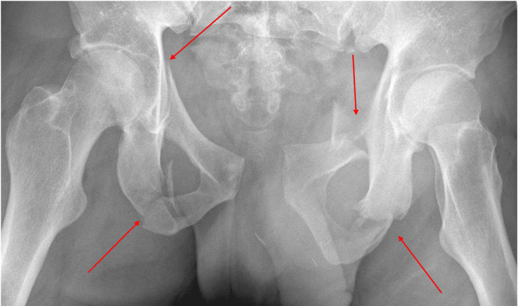
Figure 2. Pelvis radiograph.
A single frontal radiograph of the pelvis was obtained. Markedly displaced fractures were noted through the bilateral superior and inferior pubic rami with gross distraction of the pubic rami sacroiliac joint. Distraction was also suspected, with fractures extending through the S3 vertebral body. Points of fracture are indicated with red arrows.

