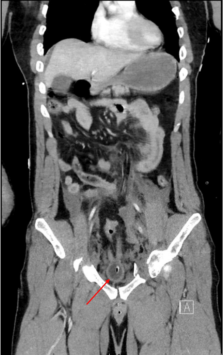
Figure 6. Abdominal CT scan highlighting the bladder.
Coronal abdominal CT scan revealing an extraordinarily compressed bladder, which could have been the result of a potential bladder rupture, especially in the context of a pelvic crush injury or open-book pelvic fracture. The compression of the bladder suggests that it has been subjected to substantial external pressure, likely from displaced pelvic bones or direct traumatic forces. This degree of compression increases the risk of bladder wall rupture. Furthermore, the presence of free fluid in the retroperitoneal space on the CT scan supports the diagnosis of a ruptured bladder, as urine may leak into this space following a tear in the bladder wall.
Note: The bladder is indicated by a red arrow.

