
Figure 2.
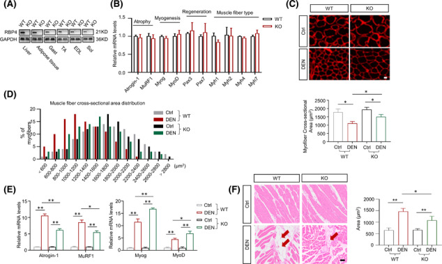
Knockout of RBP4 attenuates denervation‐induced skeletal muscle atrophy. RBP4 knockout mice were applied and subjected to denervation procedure. (A) RBP4 expression in different tissues of wild‐type and knockout mice. (B) Expression of genes related to muscle atrophy, myogenesis, muscle regeneration, and muscle fibre‐type transformation in gastrocnemius. (C) Representative immunofluorescence staining of myofibre with laminin (red) in gastrocnemius (upper) and myofibre cross‐sectional area of gastrocnemius (lower). Scale bar = 50 μm. (D) Distribution of myofibre with different cross‐sectional area in gastrocnemius. (E) The mRNA levels of muscle atrophy marker Atrogin‐1 and MuRF1, and myogenic regulator MyoD and MyoG in gastrocnemius. (F) Representative haematoxylin–eosin staining of myofibre cross‐section of gastrocnemius (left) and area of infiltrated fat in gastrocnemius (right). Arrow: Infiltrated fat. Scale bar = 50 μm. n = 8 per group, Ctrl, control; DEN, denervation; EDL, extensor digitorum longus; Gast, gastrocnemius; KO, knockout; Sol, soleus muscle; TA, tibialis anterior muscle; WT, wild type. For Figure 2 B, Mann–Whitney U tests were used. For Figure 2 C,E,F, two‐way ANOVA analyses with post‐hoc correlation were used. *P < 0.05; **P < 0.01.