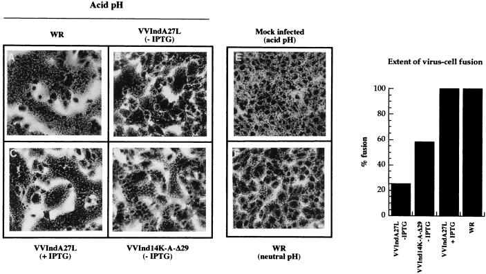
FIG. 4.
Fusion-from-without experiments at an acid pH with sucrose gradient-purified viruses. Monolayers of BSC40 cells were infected with WR (A), VVIndA27L grown in the absence (B) or presence (C) of IPTG, and VVInd14K-A-Δ29 grown without IPTG (D) under the conditions described in Materials and Methods. No cell fusion was observed in mock-infected cells at an acid pH (E) or in WR-infected cells at a neutral pH (F). The extent of virus-cell fusion was quantified by phase-contrast microscopy after counting about 103 cells from various fields (graph at right).

