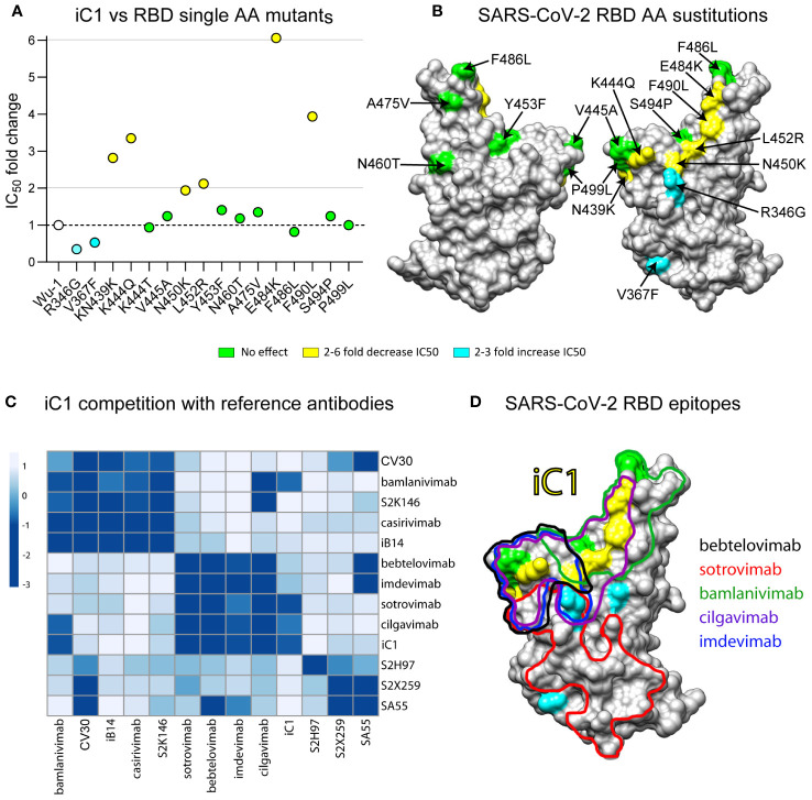
Figure 2.
Epitope mapping of iC1 binding to the RBD of SARS-CoV-2. Neutralizing activity on a panel of 15 pseudoviruses with the Spike protein (Wuhan-1) variant carrying single amino acid substitutions in the RBD region (A). A 3D model of the RBD (SARS-CoV-2) with highlighted substitutions causing effect on iC1 neutralizing activity (yellow/blue) (B). BLI analysis of iC1 competition with 12 reference antibodies with known epitope structures. Negative values (dark blue color) mean that there is a competition, positive values (white/light blue color) means that there is no competition (C). An overlay of the epitopes for the reference antibodies with single amino acid residues influencing the iC1 neutralizing activity (D).

