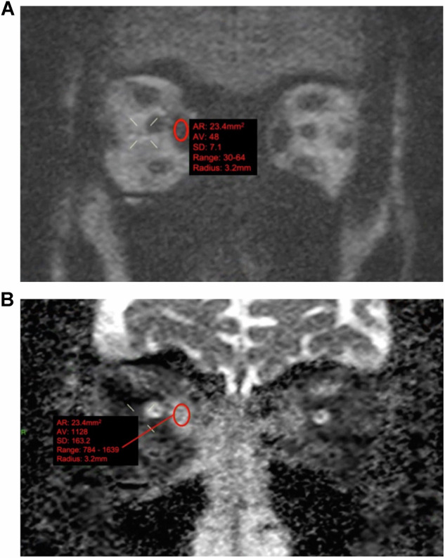
Fig. 1.

Representative examples of non-echoplanar diffusion-weighted imaging (non-EPI-DWI) of orbital EOMs in a patient with TED. Coronal orbital/EOM non-EPI-DWI MRI scan at a b value of 1000 (A) and apparent diffusion coefficient (ADC) image (B) show moderate-to-marked enlargement of the extraocular muscles and increased ADC values, notably at the right medial rectus (ADC value = 1128). Contour representing the freehand region-of-interest outline within the inner border of the active EOM
